
"Preparing ¹⁵N-Labeled LptH for Fragment-Based Drug Discovery (FBDD)" by Hayden Tucker
Updated: 1 day ago
Preparing ¹⁵N-Labeled LptH for Fragment-Based Drug Discovery (FBDD)
Hayden Tucker, Thiel College
Co-author:
Chih-Chia Su, Case Western Reserve University

Abstract
Pseudomonas aeruginosa, a Gram-negative bacterium, relies on the transport of lipopolysaccharide (LPS) across its cell envelope for survival and virulence, and carbapenem-resistant strains are now classified as a critical-priority threat for new antibiotic development. The lipopolysaccharide transport (Lpt) system moves LPS from the inner membrane to the cell surface, and the periplasmic protein LptH bridges the inner- and outer-membrane components of this pathway. Because disrupting the LptH–LptC protein–protein interaction (PPI) abolishes bacterial growth, LptH is a promising antibiotic target. This study aimed to express and purify isotopically ¹⁵N-labeled LptH suitable for fragment-based drug discovery (FBDD) using nuclear magnetic resonance (NMR) screening. His-tagged LptH was overexpressed in Escherichia coli BL21(DE3) in ¹⁵N M9 minimal medium and purified to high purity by nickel-nitrilotriacetic acid (Ni-NTA) affinity chromatography, as confirmed by sodium dodecyl sulfate–polyacrylamide gel electrophoresis (SDS-PAGE). However, extensive precipitation during dialysis reduced the final yield to 0.05 mg/mL. Substituting a Twin-Strep tag and dialyzing into phosphate-buffered saline (PBS) improved solubility and stability, yielding 1.2 mg/mL. Switching the host to E. coli C43(DE3) did not improve post-induction growth or yield. Size-exclusion chromatography (SEC) and SDS-PAGE showed that Strep-tagged LptH had superior purity, monodispersity, and durability under freeze–thaw, room-temperature, and dimethyl sulfoxide (DMSO) conditions. These findings identify the Twin-Strep construct with PBS dialysis in BL21(DE3) as the optimal route to stable, ¹⁵N-labeled LptH for downstream NMR-based fragment screening.
Introduction
Antimicrobial resistance (AMR) is among the most urgent threats to global public health. An estimated 4.95 million deaths were associated with bacterial AMR in 2019, including 1.27 million deaths directly attributable to resistant infections (Murray et al., 2022). To direct research toward the most dangerous organisms, the World Health Organization published a priority list of antibiotic-resistant bacteria, ranking carbapenem-resistant Pseudomonas aeruginosa among its critical-priority pathogens (Tacconelli et al., 2018). Because the pipeline of new antibiotics targeting Gram-negative bacteria has largely stalled, there is a pressing need for novel drug targets and discovery strategies.
Pseudomonas is a genus of Gram-negative bacteria whose metabolic diversity allows it to colonize a wide range of niches and hosts, and P. aeruginosa is a common opportunistic pathogen in immunocompromised patients, where untreated infections can become life-threatening (Huszczynski et al., 2019). A defining feature of Gram-negative bacteria is the outer membrane, whose outer leaflet is composed of lipopolysaccharide (LPS). LPS forms a selective permeability barrier that protects the cell from antibiotics, detergents, and host immune defenses, and it is a key contributor to virulence (Huszczynski et al., 2019).
LPS is synthesized at the inner membrane and must be transported across the aqueous periplasm to the cell surface. This is accomplished by the lipopolysaccharide transport (Lpt) system, a trans-envelope complex of seven proteins (LptA–LptG in Escherichia coli; the periplasmic bridging protein is designated LptH in P. aeruginosa) that forms a continuous protein bridge spanning the inner membrane, periplasm, and outer membrane (Wilson & Ruiz, 2021). LptH is the periplasmic component that physically links the inner-membrane extraction machinery to the outer-membrane translocon, making it a central, non-redundant node in the pathway.
The LptH–LptC protein–protein interaction (PPI) is essential: mutational analyses have shown that disrupting key oligomerization and interaction sites of LptH impairs LPS transport and abolishes Pseudomonas growth (Scala et al., 2020). A small molecule that binds LptH and blocks this interaction would therefore be expected to act as a potent, mechanistically novel antibiotic. However, PPI interfaces are notoriously difficult to drug with conventional high-throughput screening because they are large, flat, and lack deep binding pockets.
Fragment-based drug discovery (FBDD) is well suited to such challenging targets. Rather than screening large, drug-sized molecules, FBDD screens small, low-molecular-weight “fragments” that bind weakly but efficiently to a protein surface. Promising fragments are then grown or linked into higher-affinity leads. Over the past two decades, FBDD has matured into a mainstream strategy that has produced multiple clinical candidates and approved drugs, particularly for targets considered difficult to drug (Erlanson et al., 2016). A common way to detect weak fragment binding is solution nuclear magnetic resonance (NMR) spectroscopy, which requires the target protein to be isotopically labeled, typically with nitrogen-15 (¹⁵N), so that shifts in the protein’s NMR spectrum report on fragment binding. This same approach has been used successfully to identify fragment inhibitors of other antibiotic-resistance targets, such as the NDM-1 β-lactamase (Caburet et al., 2022).
Producing NMR-quality protein is itself a significant technical hurdle: the target must be expressed with high isotopic incorporation, purified to homogeneity, and remain soluble and stable at the concentrations, temperatures, and solvent conditions (including dimethyl sulfoxide, DMSO) used during fragment screening. The purpose of this study was therefore to establish a reproducible protocol for expressing and purifying stable ¹⁵N-labeled LptH suitable for NMR-based FBDD. Specifically, we asked which affinity tag, buffer, and host strain would yield soluble, monodisperse, isotopically labeled LptH that withstands the conditions required for fragment screening. We hypothesized that the choice of purification tag and dialysis buffer, rather than the isotopic label itself, would be the primary determinant of the protein’s solubility and stability.
Materials
Bacterial strains were E. coli BL21(DE3) and E. coli C43(DE3) chemically competent cells (obtained from standard commercial suppliers). The two LptH expression plasmids, a His-tagged construct in pET-24 (kanamycin resistance) and a Twin-Strep-tagged construct in pET-15b (ampicillin resistance), were derived from the Novagen pET vector series and maintained as laboratory stocks. Isotopic labeling was achieved using ¹⁵N M9 minimal medium prepared with ¹⁵N-ammonium chloride (Cambridge Isotope Laboratories) as the sole nitrogen source; lysogeny broth (LB) and SOC medium were used for starter and recovery cultures, respectively.
Antibiotics were kanamycin (50 µg/mL) and ampicillin (100 µg/mL), each prepared as a filter-sterilized stock in sterile deionized water. Protein expression was induced with isopropyl β-D-1-thiogalactopyranoside (IPTG) dissolved in sterile deionized water. Lysis reagents included phenylmethylsulfonyl fluoride (PMSF; a serine-protease inhibitor prepared as a stock in isopropanol) and DNase I. Chemical reagents were of molecular-biology or reagent grade and were obtained from standard commercial suppliers (e.g., MilliporeSigma and GoldBio).
For His-tagged purification, the following buffers were used, all based on 300 mM NaCl and 20 mM Tris-HCl (pH 7.5): lysis buffer (base buffer only); wash buffer 1 (base plus 20 mM imidazole); wash buffer 2 (base plus 35 mM imidazole); elution buffer (base plus 250 mM imidazole); and dialysis/size-exclusion chromatography (SEC) buffer (base plus 10% glycerol). For Strep-tagged purification, the dialysis buffer was phosphate-buffered saline (PBS; 20 mM phosphate, 100 mM NaCl, pH 7.4). Purification used gravity nickel-nitrilotriacetic acid (Ni-NTA) affinity resin (Qiagen; 2 mL packed volume) and Strep-Tactin resin (IBA Lifesciences), a 3,500 molecular-weight-cut-off (MWCO) dialysis membrane, and 10,000 MWCO centrifugal concentrators (Amicon Ultra, MilliporeSigma). Analytical SEC was performed on a Superdex 200 column (Cytiva, formerly GE Healthcare). Cells were lysed with a microfluidizer (Microfluidics), and protein concentrations were measured on a microvolume UV–Vis spectrophotometer (NanoDrop, Thermo Fisher Scientific).
Materials and Methods
Transformation and Expression of His-Tagged LptH in BL21(DE3)
To isolate LptH from the larger Lpt trans-envelope complex, the protein was overexpressed in E. coli. Selection plates were prepared from LB, agar, and 50 µg/mL kanamycin. BL21(DE3) competent cells were combined with 1 µL of a 10 ng/µL His-tagged LptH plasmid (pET-24), heat-shocked for 30 s at 42 °C, cooled on ice for 2 min, mixed with 750 µL of SOC medium, and incubated with shaking at 37 °C for 30 min. Cells were then plated in 150 µL increments and incubated inverted overnight at 37 °C.
A 250 mL flask containing 50 mL LB and 50 µg/mL kanamycin was inoculated with five colonies from the transformation plate and incubated overnight at 20 °C and 100 rpm. Two 2.8 L Fernbach flasks, each containing 1 L of ¹⁵N M9 medium with 50 µg/mL kanamycin, were each inoculated with 10 mL of the overnight starter culture. Large-scale cultures were incubated at 37 °C, and the optical density at 600 nm (OD₆₀₀) was monitored until it reached ~1.0. Cultures were then induced with IPTG (final concentration 1 mM) and grown overnight to a target OD₆₀₀ of ~2.5. Cells were harvested by centrifugation (6,000 rpm, 4 °C, 15 min) and stored at −80 °C.
Purification of His-Tagged LptH by Ni-NTA Affinity Chromatography
Cell pellets were thawed in lysis buffer on ice supplemented with 1 mM PMSF and 10 µg/mL DNase I. Cells were lysed with a microfluidizer at 15,000 psi, passing the lysate through 3–5 times for complete disruption, and clarified by centrifugation (36,000 rpm, 4 °C, 60 min; Ti-45 rotor). The supernatant was passed through a 0.45 µm membrane filter before column loading.
The clarified lysate was applied by gravity flow to 2 mL of Ni-NTA resin pre-equilibrated with wash buffer 1. The resin was washed sequentially with 20 mL of wash buffer 1 (20 mM imidazole) and 10 mL of wash buffer 2 (35 mM imidazole), and His-tagged LptH was eluted with 15 mL of elution buffer (250 mM imidazole), collecting 1.5 mL fractions. Fractions were analyzed by SDS-PAGE, and target-containing fractions were pooled, transferred to a 3,500 MWCO dialysis membrane, and dialyzed against SEC buffer (300 mM NaCl, 20 mM Tris-HCl, pH 7.5, 10% glycerol) overnight. Dialysis was required to exchange the buffer and remove nitrogen-containing compounds (e.g., imidazole) that would otherwise interfere with NMR detection of ¹⁵N-labeled protein. For SEC, pooled fractions were alternatively concentrated to ~500 µL using a 10,000 MWCO centrifugal filter and resolved on a Superdex 200 column at 0.5–1.0 mL/min. The purified protein was assessed by microvolume UV–Vis spectrophotometry, flash-frozen in liquid nitrogen, and stored at −80 °C.
Purification of Twin-Strep-Tagged LptH
After the His-tagged ¹⁵N-labeled protein was found to precipitate substantially during dialysis, the procedure was repeated with three modifications: (1) a Twin-Strep-tagged LptH plasmid (pET-15b) replaced the His-tagged pET-24 construct; (2) 100 µg/mL ampicillin replaced kanamycin for selection; and (3) the dialysis buffer was changed from SEC buffer to PBS (20 mM phosphate, 100 mM NaCl). Twin-Strep-tagged LptH was purified on a Strep-Tactin affinity column. Because the elution buffer contained desthiobiotin, pooled eluate was buffer-exchanged and concentrated in a 10,000 NMWL centrifugal filter at 3,000 × g to remove desthiobiotin and other low-molecular-weight species before downstream analysis.
Expression of LptH in E. coli C43(DE3)
Although BL21(DE3) supported successful expression of both the His-tagged and Strep-tagged constructs, cultures were observed to stop growing after IPTG induction, lowering the final yield. To test whether an alternative host would improve post-induction growth, the transformation and expression procedure was repeated using E. coli C43(DE3), a strain selected for its ability to continue growing after induction of potentially toxic membrane-associated proteins.
Results
His-tagged ¹⁵N-LptH was expressed at large scale and purified to high purity. SDS-PAGE of the Ni-NTA elution fractions showed a single dominant band at approximately 19 kDa, consistent with monomeric LptH, with little streaking or higher-molecular-weight contamination across the elution lanes (Figure 1). By SEC, this preparation eluted as a single, somewhat asymmetric peak at ~14.4 mL with a pronounced tail, indicating a predominantly monomeric species accompanied by some heterogeneity (Figure 6). Although Ni-NTA purification yielded highly pure protein, the mandatory dialysis step to remove NMR-incompatible, nitrogen-containing buffer components caused the majority of the protein to precipitate, leaving a final concentration of only 0.05 mg/mL.
Several factors could have contributed to this precipitation, prompting additional trials to test alternative tags, buffers, and host strains. Replacing the His tag with a Twin-Strep tag was undertaken first, both to enable an alternative, highly selective purification chemistry (Strep-Tactin) and to test a different dialysis buffer. Strep-Tactin purification produced a highly pure preparation: SDS-PAGE showed a dominant band at ~22–24 kDa, slightly higher than the His-tagged form because of the added tag mass (Figure 2).
Dialyzing the Strep-tagged construct into PBS resulted in little to no visible precipitation and a substantially higher final yield of 1.2 mg/mL, a roughly 24-fold improvement over the His-tagged, SEC-buffer condition. Analytical SEC confirmed the quality of this material: Strep-tagged LptH eluted as a sharp, symmetric peak at ~15.0 mL with only a minor secondary peak, indicating greater monodispersity than the His-tagged preparation (Figure 7). To confirm that the observed loss was tag/buffer-dependent rather than intrinsic to LptH, wild-type (untagged) LptH was also purified and showed a clear ~19 kDa product with no visible precipitation during dialysis, establishing a solubility benchmark (Figure 3).
To determine whether the isotopic label itself altered behavior, unlabeled and ¹⁵N-labeled His-tagged LptH were compared by SEC. Both eluted at the same volume (~15 mL); the ¹⁵N-labeled sample showed a lower peak amplitude (~7 versus ~23 mAU), reflecting reduced yield rather than any change in elution position or oligomeric state (Figure 9). This supported the interpretation that ¹⁵N labeling does not compromise solubility, and that the tag and dialysis buffer were the operative variables.
Because FBDD by NMR requires the target to remain folded and monodisperse at room temperature and in the presence of DMSO, the stability of Strep-tagged LptH was assessed under these conditions. A direct overlay of His- and Strep-tagged material showed comparable single-peak elution near 15 mL, with the Strep-tagged construct retaining a clean profile (Figure 8). Strep-tagged LptH also retained a consistent ~15 mL peak after 24 h at room temperature and after 24 h in DMSO (Figure 10), and near-identical elution profiles were obtained when the room-temperature and DMSO-incubated samples were compared directly (Figure 11). Finally, samples held for 24 h in DMSO versus water gave overlapping main peaks (Figure 12), confirming that the protein tolerates the inert screening solvent used in fragment campaigns.
For structural context, a cryo-electron microscopy (cryo-EM) reference image of the Lpt trans-envelope bridge is shown in Figure 5. In contrast to these successes, switching the expression host from BL21(DE3) to C43(DE3) did not improve outcomes: SDS-PAGE of the C43(DE3) Strep-tagged preparation showed abundant protein in the flow-through and wash fractions but only a faint target band in the elution fractions, indicating low recovery relative to BL21(DE3) (Figure 4).
Discussion
The successful expression and purification of LptH in E. coli confirms that this common host is well suited for producing the periplasmic bridging protein of the Lpt system. The high purity achieved by a single Ni-NTA step (Figure 1) shows that the affinity-tag strategy efficiently separates LptH from the complex mixture of host proteins present after lysis. The central obstacle was not expression or purity but solubility during buffer exchange.
The most consequential finding was that the majority of His-tagged ¹⁵N-LptH precipitated during dialysis into SEC buffer, whereas wild-type LptH (Figure 3) and Twin-Strep-tagged LptH dialyzed into PBS (Figures 2 and 7) remained soluble. Because the ¹⁵N label had no effect on elution behavior (Figure 9), and because isotopic substitution is not expected to alter a protein’s solubility, the label can be excluded as the cause. The evidence instead points to the combination of affinity tag and buffer as the determining factors, consistent with our hypothesis. This interpretation aligns with the established observation that Strep-Tactin purification yields high-purity target protein under mild, physiological conditions, which can favor solubility relative to imidazole-based elution and exchange (IBA Lifesciences, 2015; Thermo Fisher Scientific, n.d.). The precise mechanism, whether PBS better supports LptH’s surface charge and hydration than the SEC buffer, or whether the His tag itself promotes aggregation under these conditions, remains to be determined and warrants systematic buffer screening (for example, varying pH, ionic strength, and additives).
The stability results are directly relevant to the downstream application. Fragment screening by NMR requires a target that remains folded, monomeric, and soluble at room temperature for the duration of a screen and that tolerates DMSO, in which fragment libraries are dissolved. The SEC overlays demonstrate that Strep-tagged LptH satisfies these requirements: it retained a single, consistent elution peak after freeze–thaw, after 24 h at room temperature, and after 24 h in DMSO (Figures 8, 10, 11, and 12). Establishing this stability window is a prerequisite for any FBDD campaign and mirrors the sample-quality standards used in fragment screens against other antibiotic-resistance targets, such as NDM-1 β-lactamase (Caburet et al., 2022; Erlanson et al., 2016).
The attempt to increase yield by switching to E. coli C43(DE3) was unsuccessful; recovery was lower than in BL21(DE3) (Figure 4), indicating no advantage for this construct under the conditions tested. The growth arrest observed after IPTG induction in BL21(DE3) suggests that LptH overexpression imposes a metabolic or membrane-associated burden on the host. Future work could address this through lower induction temperatures, reduced IPTG concentrations, auto-induction media, or tightly regulated expression systems to extend post-induction growth and improve isotopic yield.
This study has limitations. Yields are reported from single representative preparations rather than biological replicates, and the mechanism of His-tag/SEC-buffer precipitation was not resolved. Protein identity was inferred from migration and elution behavior; confirmatory methods such as mass spectrometry or Western blotting, and a folding readout such as a one-dimensional or ¹⁵N-HSQC NMR spectrum, would strengthen the characterization. Nonetheless, the consistent, convergent behavior of the wild-type and Strep-tagged constructs provides a coherent and internally consistent picture of the variables that govern LptH solubility.
Conclusion
The goal of preparing stable, ¹⁵N-labeled LptH for fragment-based drug discovery was achieved through a series of expression and purification trials that progressively refined the protocol. The His-tagged construct yielded highly pure protein but precipitated extensively during dialysis, reducing yield to 0.05 mg/mL. Substituting a Twin-Strep tag and dialyzing into PBS prevented precipitation and increased the yield to 1.2 mg/mL, while comparison with wild-type LptH and unlabeled controls demonstrated that the isotopic label was not responsible for the loss. Switching from E. coli BL21(DE3) to C43(DE3) did not improve viability or yield after induction. Stability trials confirmed that Strep-tagged LptH retained structural integrity after freeze–thaw, 24 h at room temperature, and exposure to DMSO, conditions required for NMR-based screening. Together, these results identify the Twin-Strep tag, PBS dialysis, and BL21(DE3) host as an effective route to high-quality, ¹⁵N-labeled LptH, laying the groundwork for future NMR-based FBDD campaigns targeting the essential LptH–LptC interaction in Pseudomonas and, ultimately, the development of urgently needed antibiotics against Gram-negative infections.
Figure Legends
Legends are listed below in order.
Figure 1. Ni-NTA elution of ¹⁵N-His-tagged LptH resolved by SDS-PAGE. Lane M, molecular-weight marker (kDa); lanes 1–14, sequential Ni-NTA elution fractions. A single dominant band migrates at ~19 kDa (monomeric LptH) across lanes 2–14, with minimal higher-molecular-weight contamination, indicating high purity.

Figure 2. Strep-Tactin elution of ¹⁵N-Twin-Strep-tagged LptH resolved by SDS-PAGE. Lane M, marker; the adjacent reference lane is labeled “LptH Liposol,” followed by lanes 1–10 of sequential Strep-Tactin elution fractions. The tagged construct migrates at ~22–24 kDa, higher than the His-tagged form because of the added Twin-Strep tag, with a faint minor band near 17 kDa.
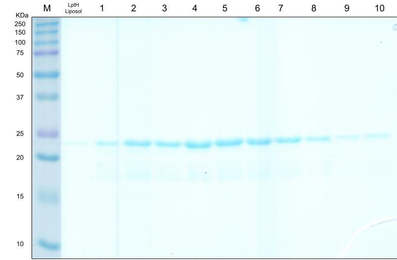
Figure 3. Purification of wild-type (untagged) LptH resolved by SDS-PAGE. Lane M, marker; a combined flow-through/wash lane; lanes 1–10, elution fractions. A ~19 kDa product appears in the central fractions (lanes 3–7). Wild-type LptH showed no visible precipitation during dialysis and serves as a solubility benchmark.

Figure 4. Expression and purification of ¹⁵N-Twin-Strep-tagged LptH in E. coli C43(DE3), SDS-PAGE. Lane M, marker; flow-through and wash 1 lanes contain abundant total protein, while only a faint target band (~22 kDa) appears in elution lanes 3–7, indicating low recovery relative to BL21(DE3).

Figure 5. Original cryo-electron microscopy (cryo-EM) micrograph of the Lpt trans-envelope bridge. This micrograph, acquired as part of this study, shows the Lpt trans-envelope bridge in the context of the bacterial membrane; the boxed region and arrow indicate the feature of interest.

Figure 6. Size-exclusion chromatography (SEC) of His-tagged LptH (Superdex 200). UV absorbance (mAU) versus elution volume (mL). A single asymmetric peak elutes at ~14.4 mL with a pronounced tail, consistent with a predominantly monomeric species showing some heterogeneity.

Figure 7. SEC of Twin-Strep-tagged LptH (Superdex 200). A sharp, symmetric main peak elutes at ~15.0 mL with a minor peak near 19.2 mL, indicating higher monodispersity and purity than the His-tagged preparation (compare Figure 6).

Figure 8. Overlaid SEC traces comparing His-tagged and Strep-tagged LptH integrity. Both constructs elute as a single peak near 15 mL, indicating comparable elution behavior and retained structural integrity after freezing.

Figure 9. Overlaid SEC traces comparing unlabeled and ¹⁵N-labeled His-tagged LptH. Both samples elute at ~15 mL; the ¹⁵N-labeled preparation (orange) shows a lower peak amplitude (~7 vs. ~23 mAU), reflecting reduced yield rather than a change in elution position, indicating that isotopic labeling does not alter the oligomeric state.

Figure 10. Overlaid SEC traces assessing Strep-tagged LptH stability. Freshly purified Strep-tagged LptH, the same sample after 24 h at room temperature, and after 24 h in DMSO all share a common peak at ~15 mL, indicating the protein remains folded and monodisperse under FBDD-relevant conditions.

Figure 11. Overlaid SEC traces comparing Strep-tagged LptH after 24 h at room temperature versus 24 h in DMSO. Near-identical elution profiles indicate that the protein tolerates prolonged DMSO exposure.
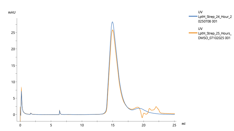
Figure 12. Overlaid SEC traces comparing Strep-tagged LptH held 24 h in DMSO versus 24 h in water. Overlapping main peaks at ~15 mL confirm buffer/solvent stability under both conditions.

References
Caburet, J., Boucherle, B., Bourdillon, S., Simoncelli, G., Verdirosa, F., Docquier, J.-D., Moreau, Y., Krimm, I., Crouzy, S., & Peuchmaur, M. (2022). A fragment-based drug discovery strategy applied to the identification of NDM-1 β-lactamase inhibitors. European Journal of Medicinal Chemistry, 240, 114599. https://doi.org/10.1016/j.ejmech.2022.114599
Erlanson, D. A., Fesik, S. W., Hubbard, R. E., Jahnke, W., & Jhoti, H. (2016). Twenty years on: The impact of fragments on drug discovery. Nature Reviews Drug Discovery, 15(9), 605–619. https://doi.org/10.1038/nrd.2016.109
Huszczynski, S. M., Lam, J. S., & Khursigara, C. M. (2019). The role of Pseudomonas aeruginosa lipopolysaccharide in bacterial pathogenesis and physiology. Pathogens, 9(1), 6. https://doi.org/10.3390/pathogens9010006
IBA Lifesciences. (2015). Protein purification with Strep-Tactin. https://www.iba-lifesciences.com/strep-tag/strep-tactin/
Murray, C. J. L., Ikuta, K. S., Sharara, F., Swetschinski, L., Robles Aguilar, G., Gray, A., … Naghavi, M. (2022). Global burden of bacterial antimicrobial resistance in 2019: A systematic analysis. The Lancet, 399(10325), 629–655. https://doi.org/10.1016/S0140-6736(21)02724-0
Scala, R., Di Matteo, A., Coluccia, A., Lo Sciuto, A., Federici, L., Travaglini-Allocatelli, C., Visca, P., Silvestri, R., & Imperi, F. (2020). Mutational analysis of the essential lipopolysaccharide-transport protein LptH of Pseudomonas aeruginosa to uncover critical oligomerization sites. Scientific Reports, 10(1), 11928. https://doi.org/10.1038/s41598-020-68054-7
Tacconelli, E., Carrara, E., Savoldi, A., Harbarth, S., Mendelson, M., Monnet, D. L., … Zorzet, A. (2018). Discovery, research, and development of new antibiotics: The WHO priority list of antibiotic-resistant bacteria and tuberculosis. The Lancet Infectious Diseases, 18(3), 318–327. https://doi.org/10.1016/S1473-3099(17)30753-3
Thermo Fisher Scientific. (n.d.). Ni-NTA purification system: For purification of polyhistidine-containing recombinant proteins. https://tools.thermofisher.com/content/sfs/manuals/ninta_system_man.pdf
Wilson, A., & Ruiz, N. (2021). Transport of lipopolysaccharides and phospholipids to the outer membrane. Current Opinion in Microbiology, 60, 51–57. https://doi.org/10.1016/j.mib.2021.01.006


Comments